


WOMAN HEALTH
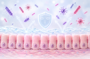
Woman health relies on the proper function of the vaginal epithelium, which serves as the first line of defense against pathogens while maintaining tissue homeostasis, mucosal protection, and balanced immune responses.
To support the development of innovative products for woman health, we have established a comprehensive portfolio of cell-based assays that enables the evaluation of multiple mechanisms of action, including anti-adhesion and anti-infective activity against vaginal pathogens, stimulation of mucus production, modulation of cytokine release, epithelial barrier protection, and other functional markers relevant to vaginal health.

Analyse Your Product's Impact On Woman's Health

We assess the beneficial properties of your product(s) on woman health by analysing its effec on:


ANTI-ADHESION ASSAY
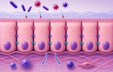
The anti-adhesion assay evaluates a product’s ability to prevent pathogens from attaching to host vaginal and/or cervical epithelial cells.
The assay can be performed as an exclusion, competition, or displacement assay.

Vaginal / Cervical Cell lines
Incubation with
product
Aerobe
Anaerobe
Microaerophile
Incubation with
pathogen
Quantification
Fluorescence
Bioinformatic & customized visualization
CFU determination
The Assay
This assay, differentiated host cells are incubated with the selected pathogen(s) and the test compound(s), such as a probiotic, postbiotic, or plant extract. The number of pathogens that successfully adhere to the cells is then quantified by determining CFUs and/or AFUs.
Comparison of CFUs/AFUs in the presence and absence of the test product allows the identification of potential anti-adhesion properties.
The assay can be performed as an exclusion, competition, or displacement assay.

MUCUS PRODUCTION
The vaginal mucus layer forms the first line of defense against invading pathogens. It acts as a physical and biochemical barrier that traps microorganisms, limits their access to the epithelial surface, and contains antimicrobial molecules that contribute to innate mucosal immunity. In addition, vaginal mucus helps maintain tissue hydration, lubrication, and overall mucosal homeostasis.
The Assay
This assay evaluates the ability of products to to stimulate or restore mucus production therefore provides valuable insight into its potential to enhance the natural protective function of the vaginal mucosa.
In this assay, vaginal and/or cervical epithelial cells are cultured and treated with the test product. After incubation, secreted mucus can be staining and/or specific ELISAs. In parallel, muc-genes expression can be assessed by qRT-PCR.

Cell differentiation
Add the test product
Incubation with test product
qRT-PCR
quantification
Bioinformatic & customized visualization
Staining or ELISA
CYTOKINE PRODUCTION
The vaginal epithelium actively contributes to mucosal immunity through the production of cytokines and chemokines in response to pathogens and external stimuli. These signaling molecules regulate inflammation, coordinate immune cell recruitment, and play a key role in protecting the vaginal mucosa against infection while maintaining tissue homeostasis.

Eventual
pre-treatment
Quantification of cytokine expession
The Assay
This assay evaluates the ability of products to modulate cytokine production, including the stimulation of protective immune responses or the reduction of inflammatory signaling. It therefore provides valuable insight into their potential to support the natural protective function of the vaginal mucosa.
In this assay, vaginal epithelial cells are cultured and treated with the test product. After incubation, secreted cytokines can be quantified using specific ELISAs. In parallel, cytokine gene expression can be assessed by qRT-PCR.
The assay can be performed under physiological conditions, as well as under simulated inflammatory or infectious conditions.
Differentiated cells
Incubation with product
Bioinformatic & customized visualization
Quantification of cytokine secretion

EXTENDED IMMUNOMODULATORY PROPERTIES
The vaginal epithelium and resident immune cells closely interact to maintain mucosal homeostasis and provide an effective defense against pathogens. Upon exposure to microorganisms or bioactive compounds, epithelial cells secrete cytokines and chemokines that regulate the activation and recruitment of immune cells, while immune cells further shape epithelial responses through reciprocal signaling.
Our VK2/E6E7–THP-1 or VK2/E6E7–CD14⁺ co-culture models closely mimic this epithelial–immune cell crosstalk, enabling the evaluation of the immunomodulatory properties of probiotics, postbiotics, plant extracts, pharmaceuticals, and other bioactive compounds under physiological, inflammatory, or infectious conditions.
The Assay
This assay aims at analysing the immune modulatory effect of product(s) in the vaginal context.
The VK2/E6E7/THP-1 o VK2/E6E7/CD4+ co-culture model mimics the interaction between vaginal epithelial cells and subepithelial immune cells, thereby reproducing key aspects of the immune response to intestinal compounds.
To characterize the immunomodulatory properties of test products, the said products are applied to the co-culture and their effect on cytokine secretion (via ELISA) and/or gene expression (via qRT-PCR). are quantified.
Differentiated
immune cells

Differentiated
epithelial cells
Incubation with product
Bioinformatic & customized visualization
Quantification of cytokine secretion
Eventual
pre-treatment
Quantification of cytokine expression
BARRIER INTEGRITY
The vaginal epithelium forms a highly specialized physical barrier that protects the underlying tissue from invading pathogens while maintaining a controlled exchange of nutrients and signaling molecules. This barrier is maintained by tight intercellular junctions and a well-organized epithelial layer, both of which are essential for preventing microbial penetration and preserving mucosal homeostasis.
Assessment of vaginal barrier integrity provides valuable insight into the ability of a product to protect or restore epithelial function under physiological, inflammatory, or infectious conditions.
Keratinocytes (HaCaT) &
Dermal fibroblasts (NHDF)
Incubation with the product(s)
UV treatment
qRT-PCR
for specific markers
Bioinformatic & customized visualization
Quantification of markers

The Assay
This assay evaluates the ability of a test compound to maintain or restore vaginal epithelial barrier integrity.
Differentiated vaginal epithelial monolayers are exposed to pre-treatment (dextran sulfate sodium (DSS) or a cytokine cocktail) to induce inflammation and disrupt barrier function.
The protective effect of the test product is quantified by transepithelial electrical resistance (TEER), paracellular permeability, and/or the expression of key junctional proteins.
The test compound can be applied before, during, or after inflammation induction (leaky gut model), depending on the experimental design and intended readout.

