


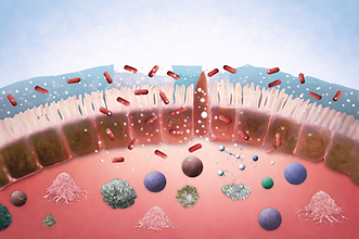
INTESTINAL BARRIER INTEGRITY
The intestinal epithelial barrier plays a vital role in maintaining health by regulating the selective passage of nutrients while preventing the entry of pathogens and harmful substances. Disruption of this barrier, often referred to as “leaky gut”, can result in low-grade chronic inflammation.
This dysfunction is implicated in a range of gastrointestinal and systemic disorders, including inflammatory bowel disease (IBD), type 1 diabetes, celiac disease, and multiple sclerosis.


PERMEABILITY
A leaking gut barrier facilitates translocation of harmful substances and the development of inflammation.
The Assay
-
This assay evaluates the ability of a test compound to maintain or restore intestinal barrier integrity.
-
Differentiated intestinal epithelial monolayers are exposed to dextran sulfate sodium (DSS) or a cytokine cocktail to induce inflammation and disrupt barrier function.
-
The protective effect of the test product is quantified by measuring Lucifer Yellow (LY) permeability and/or by analyzing the expression of tight junction markers.
-
The test compound can be applied before, during, or after inflammation induction (leaky gut model), depending on the experimental design and intended readout.
Incubation with the product

Simulation of inflammation
(DSS or cytokines)
Q-RT-PCR
for specific markers
Incubation with LY
Intestinal cells from:
-Human
-Feline
-Canine (primary cells)
-Porcine
Bioinformatic
& customized visualization
Measure
Basolateral LY
WOUND HEALING
The intestinal epithelium renews itself every 3–5 days, requiring precise regulation of cell proliferation, differentiation, and wound healing to maintain tissue integrity.

Bioinformatic
& customized visualization
Cell differentiation in insert
Removing insert
Incubation with test product
Time to close the gap
RT-PCR of markers
The Assay
-
This assay evaluates the ability of products to promote epithelial repair.
-
Intestinal epithelial cells are cultured and differentiated using the ibidi Culture-Insert 2-Well system, which creates a standardized, cell-free “wound” area. Following barrier removal, gap closure is monitored microscopically over time, providing a quantitative measure of cell migration and regeneration. Optionally, the expression of repair and proliferation markers can be analyzed by qRT-PCR.


MUCUS PRODUCTION
-
The mucus forms a protective barrier that shields the intestinal lining from pathogens, toxins, and mechanical damage while maintaining a stable environment for beneficial microbes. It also supports healthy immune responses and ensures smooth nutrient absorption by preventing inflammation and preserving epithelial integrity.
The Assay
-
The Assay evaluates a compound’s ability to stimulate mucin secretion by epithelial cells.
-
In this assay, HT29-MTX intestinal epithelial cells are cultured under semi-wet conditions with mechanical stimulation to promote mucus formation. Mucin expression can be modulated by introducing inflammatory conditions (e.g., cytokine mix).
-
The test product can be applied before, during, or after induction, depending on the desired readout. Mucus production is quantified using specific staining methods (Alcian Blue or Periodic Acid–Schiff), ELISA, or RT-qPCR for mucin gene expression.

Quantification transcription (MUC2, MUC5AC, MUC1, MUC4)
Add Test Product
Cell growth and differentiation
in semi-liquid conditions
Bioinformatic
& customized visualization
Quantification secretion (MUC2, MUC3, MUC4, MUC5AC)
ANTI OXIDATIVE STRESS
-
Mucus forms a protective barrier that shields the intestinal lining from harmful substances while supporting beneficial microbes and maintaining epithelial integrity. Oxidative stress, which occurs when reactive oxygen species overwhelm the body’s antioxidant defenses due to factors like inflammation, infection, poor diet, pollutants, or cellular metabolism, can damage intestinal cells, weaken the gut barrier, and trigger inflammation.


Add an oxidative stressor
Cell growth and differentiation
Incubate with product
Measure ROS
The Assay
This assay evaluates the ability of a test compound to protect epithelial cells against oxidative stress.
-
Epithelial cells are pre-incubated with the test product, then exposed to an oxidative stressor (e.g., H₂O₂ or AAPH) in the presence of a fluorescent probe (e.g., DCFH-DA).7
-
The probe reacts with reactive oxygen species (ROS) to generate a measurable fluorescent signal, reflecting intracellular oxidative activity.
Have a non-binding consultation with one of our experts to find out the best solution for your needs.
Contact us.

