


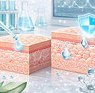
SKIN HEALTH
Healthy skin is essential for overall well-being, serving as both a protective barrier and a dynamic immunological and sensory interface.
Because skin aging and acne are major consumer concerns, we have established several cell-based assays that provide clear insights into the beneficial effects of your products on these two key skin health challenges.

Analyse Your Product's Impact On Acne
We assess the anti-acne properties of your product(s) by analysing its effect on:


Analyse Your Product's Impact On Skin Aging

We assess the anti-aging properties of your product(s) by analysing its effect on:


PERMEABILITY ASSAY
The skin epithelium serves as a critical barrier protecting the body from environmental stressors. Compromised barrier integrity can lead to inflammation, irritation, and chronic skin disorder

Keratinocyte cells
(HaCaT)
Incubation with
product
Simulation of inflammation
(EDTA, mild SDS)

Incubation with LY
qRT-PCR
for specific markers
Bioinformatic & customized visualization
Measure
basolateral LY
The Assay
This assay evaluates the barrier-protective properties of probiotics, postbiotics, or cosmetic ingredients on keratinocyte monolayers.
To simulate barrier disruption and inflammatory stress, monolayers are exposed to EDTA or SDS. The protective effect of the test product is quantified by measuring Lucifer Yellow (LY) permeability and by analyzing the expression of barrier integrity markers.

The Assay
This assay evaluates the ability of products to promote epithelial repair and regeneration.
Keratinocytes are cultured and differentiated using the ibidi Culture-Insert well system, which creates a standardized, cell-free “wound” area.
Following barrier removal, gap closure is monitored microscopically over time, providing a quantitative measure of regenerative capacity. Additionally, the expression or secretion of healing-related and extracellular matrix (ECM) markers can be quantified.
WOUND HEALING
The skin epithelium continuously renews and repairs itself to maintain an effective barrier against environmental stressors.

Remove insert
Cell differentiation in insert
Incubation with test product
qRT-PCR of markers
Time requires to close the gap
Bioinformatic & customized visualization
ANTI-APOPTOSIS
Excessive apoptosis in skin cells can compromise barrier integrity, accelerate skin aging, and impair tissue regeneration.


Bioinformatic & customized visualization
Quantification of apoptosis-related markers
Keratinocytes
Differentiation
Incubation with pro-apoptotic agent & the product
The Assay
This assay evaluates the ability of products to protect keratinocytes against apoptosis induced by chemical or oxidative stress.
Keratinocytes are exposed to pro-apoptotic stimuli in the presence or absence of the test product. The anti-apoptotic effect is quantified by measuring cell viability and/or the expression of apoptosis-related markers (i.e. Cytochrome c, Bax).

The Assay
This assay evaluates the ability of products to stimulate ECM synthesis and regulate matrix remodeling in dermal fibroblasts.
Cells are incubated with the test product under controlled conditions, and the expression or secretion of ECM components and remodeling markers (including pro-collagen I/III, fibronectin, hyaluronan, MMP-1/3/9) is quantified.
EXTRACELLULAR MATRIX
The extracellular matrix (ECM) provides essential structural and biochemical support to the skin, maintaining its firmness, elasticity, and tissue integrity. A decline in ECM synthesis or dysregulated remodeling contributes to skin aging, loss of elasticity, and impaired wound repair.

qRT-PCR
for specific markers
Dermal fibroblasts
(NHDF)
Incubation with the product(s)
Bioinformatic & customized visualization
Quantification of markers
UV PROTECTION
Exposure to ultraviolet (UV) radiation induces oxidative stress, DNA damage, and inflammation, accelerating skin aging and impairing barrier function.


Incubation with the product(s)
Bioinformatic & customized visualization
Quantification of markers
qRT-PCR
for specific markers
UV treatment
Keratinocytes (HaCaT) &
Dermal fibroblasts (NHDF)
The Assay
This assay evaluates the ability of products to protect skin cells against UV-induced damage.
Keratinocytes are exposed to UVA to assess barrier and antioxidant protection, while dermal fibroblasts are irradiated with UVB to evaluate anti-photoaging effects.
Cell viability, ROS generation (DCFDA, MitoSOX), and the expression of stress- and repair-related markers are quantified (CPDs, IL-6, IL-8, TNF-α, TSLP, pro-collagen I/III, MMP-1/3/9 etc).
ANTI-MICROBIAL
Cutibacterium acnes plays a central role in the development of acne.
The Assays
The diffenrent assays evaluating the antimicrobial and anti-adhesion properties of products against C. acnes are described in the section anti-microbial properties.
Co-culture, well diffusion, or overlay methods to assess growth inhibition and bacteriostatic or bactericidal effects of the test product.
Anti-adhesion assays to determine the ability of the test compound to prevent bacterial attachment to host cells.

Overlay & Diffusion Assays

Anti-Adhesion Assay

Co-Culture
Assay

Aggregation
Assay
HYPER-KERATINIZATION
Excessive keratinization can lead to follicular occlusion, rough skin texture, and acne formation

Keratinocytes
Incubation with the product(s)
qRT-PCR for specific markers
Bioinformatic & customized visualization
Quantification of markers

The Assay
The Assay evaluates the ability of products to modulate keratinocyte differentiation and support barrier homeostasis.
Keratinocytes are cultured under differentiation-inducing conditions in the presence or absence of the test product.
The expression of key keratinization and barrier markers (filaggrin, involucrin, loricrin, keratin 1/10) are quantified by qRT-PCR and/or immunostaining.
OXIDATIVE STRESS
Excessive Oxidative stress in sebocytes contributes to lipid peroxidation, inflammation, and acne.


Human sebocytes (SZ95)
Incubation with the product(s)
Oxidative Stress
qRT-PCR for specific markers
Bioinformatic & customized visualization
Quantification of markers
The Assay
This assay evaluates the anti-oxidative potential of products in human sebocytes (SZ95 cells).
Cells are treated with the test product and exposed to oxidative stressors under controlled conditions. Global ROS levels are quantified using the DCFDA fluorescence assay.
Additionally, MitoSOX can be employed to detect mitochondrial superoxide, while testing TMRE or JC-1 help assessing mitochondrial membrane potential (ΔΨm) integrity.

ANTI-LIPOGENESIS
Excessive sebum production is a key factor contributing to acne formation and microbial imbalance.
The Assay
This assay evaluates the ability of products to inhibit lipogenesis in human sebocytes.
Cells are treated with the test product under controlled conditions, and intracellular lipid accumulation is quantified by Nile Red neutral lipid staining, providing a direct measure of sebum synthesis.
In parallel, 5α-reductase activity, a key enzyme involved in androgen-mediated lipogenesis, is quantified to further characterize the product’s sebum-regulating potential.

Human sebocytes (SZ95)
Incubation with the product(s)
UV treatment
Staining with
Nile Red
Bioinformatic & customized visualization
Quantification

